
Arthroscopy - knee joint
Introduction
Arthroscopy of the knee joint is the most frequently performed and most sophisticated form of arthroscopic joint surgery. It employs a minimally invasive endoscopic technique which enables excellent visualization of the knee joint and also allows surgical treatment of any damage found. This minimally invasive procedure enables us to reduce postoperative tenderness, thereby accelerating postoperative rehabilitation and permitting the patient to return to his usual daily activities more rapidly.
Anesthesia
For knee arthroscopy, we employ general anesthesia or spinal anesthesia (which numbs the lower extremities by introducing anesthetic into the spinal canal), potentially combined with regional anesthesia. General anesthesia is gentle on the patient and allows for good muscle relaxation. To reduce intraoperative bleeding, we use a pneumatic tourniquet, positioned roughly at the midpoint of the thigh. Regional anesthesia is most often used to reduce postoperative pain. This involves a blockade of the femoral nerve, particularly in surgery on cruciate ligaments. The blockade lasts for 24-48 hours after surgery, substantially cutting back on the need for opiates.
Position and Access Points
During arthroscopy of the knee joint, the patient is placed in a supine position, with the leg being operated on allowed to hang over the edge of the operating table to permit manipulating it during the operation.
Surgical Access Points
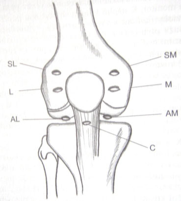
Basic:
- AL - anterolateral: This point is located approximately 1.5 cm external to the lig. patellae, 1.5 cm below the edge of the patella. We use this as an entry port for optics.
- AM - anteromedial: This point is located approximately 1.5 cm medial to the lig. patellae, 1.5 cm below the edge of the patella. We use this as an entry port for surgical instruments.
Other Access Points:
- SL - suprapatellar lateral
- SM - suprapatellar medial
- M - central patellar medial
- M - central patellar lateral
- C - central
Technological Tools
We use a relatively complex set of optical and surgical instruments for arthroscopic surgery, placed in a so-called arthroscopic tower. They include:
- Camera (analog, digital) – after attachment to the eyepiece of the arthroscope, enables image transfer to the monitor.
- Light source – xenon lamp with optical cable
- ASK pump – maintains the pressure and flow of solution through the joint
- Shaver – motorized rotary cutter
- Vulcan – used to treat soft tissue on the principle of high-frequency thermocoagulation
- Documentation equipment – printer, smart card, video, DVD, HD
We also make use of:
- Trocar – a cannula whose mandrin is replaced by the arthroscope after introduction into the joint
- Arthroscope –a rigid or flexible tube with a sapphire lens, enabling a side view of 30° or 70°
- Instruments – punch, scissors, knives, forceps, extractors, rasps, hooks
- Specialized instruments – reconstruction of ligaments, mosaic plastics, shoulder stabilization
Indications
- meniscal injury
- injury to the anterior cruciate ligament (ACL)
- cartilage injury
- hyperpression of the patella
- free bodies present in the joint
- arthrofibrosis, synovitis
- intraarticular fracture